
Fig. 1.
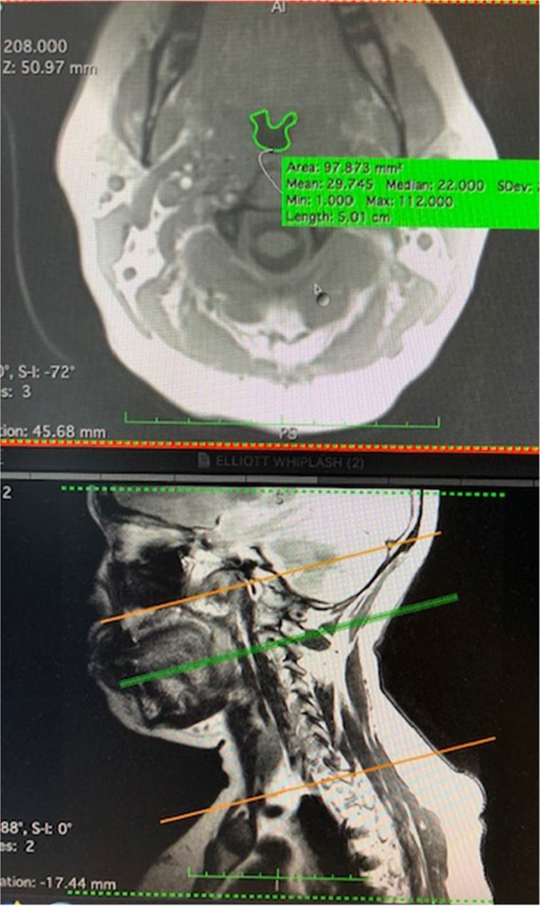
Pharyngeal contouring on MRI. This figure illustrates an axial slice of the C-spine (top image), where the green trace delineates the pharyngeal lumen. This axial slice represents C2 of the cervical spine, corresponding to the green line on the sagittal image (bottom image). The orange lines marks the region of interest from which axial slices were taken to represent the pharynx