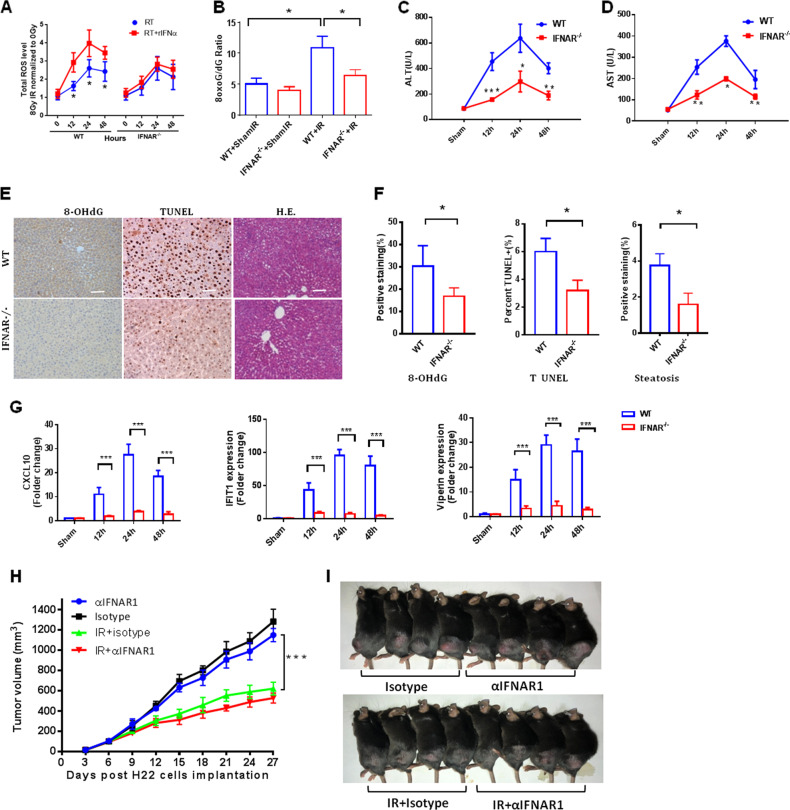
Fig. 5.
Blockade of type 1 IFN signaling protects mice from radiation-induced liver injury without affecting tumor growth. Measurement of intracellular total ROS (a) in irradiated primary WT mouse hepatocytes and IFNAR−/− cells with or without rIFN-α (n = 7) at 0, 12, 24, and 48 h postirradiation (30 Gy). b 8-oxo-G/deoxyguanosine ratios in IFNAR−/− versus WT mice 24 h after whole-liver irradiation (30 Gy IR). Serum c ALT and d AST levels in IFNAR−/− and WT mice at 12, 24, and 48 h after IR. e 8-hydroxy-2′-deoxyguanosine (8-OHdG) and TUNEL staining in the liver was determined by immunohistochemistry. Steatosis was analyzed in H&E staining. Scale bar: 100 μm. f Quantification of TUNEL-positive cells in each high-power field and positive 8-OHdG signal in a unit area, and steatosis score at the indicated region. g Expression of CXCL10, IFIT1, and viperin in IFNAR−/− mouse hepatocytes versus WT cells at 12, 24, and 48 h after irradiation. h H22 tumor size (mean ± SD, n = 7 mice/group) irradiated with 8 Gy for 3 consecutive days in mice that received intraperitoneal injection with 250 mg of a-IFNAR1 antibody (Clone MAR1-5A3, BioXCell) or matched isotype control (clone MOPC21, BioXCell) 1 day prior to whole-liver irradiation (8 Gy x 3) followed by injections every other day. Significant differences were determined using two-way ANOVA. i Representative mice with subcutaneous H22 tumors at termination, 27 days after tumor inoculation. Mean ± SD; n = 5. *P < 0.05, **P < 0.01, and ***P < 0.001

