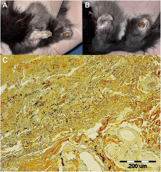
Figure 1.

Skin lesions on the right hindlimb of a pet rabbit with syphilis. (A, left) Detailed view of healthy (2nd) and syphilis affected (3rd) nail and toe. (B, right) Note the fractured and deformed claws/nails on both the 3rd and 4th toes. White scaly lesions were seen on both affected claws/nails, the eponychium, and the distal parts of terminal phalanges. (C, bottom) Wartin-Starry silver stain highlights spirochetes in dermis, magnification 200 × . Histopathological examination of the lesion in the area of claw and surrounding connective dermal tissue showed presence of proliferating fibrovascular tissue, moderate mixed inflammatory infiltrate with predominance of lymphocytes, plasma cells, and macrophages, with admixture of lesser number of neutrophils. There was superficial erosion of epithelium, areas of serocellular crusts, focally small hemorrhages and deposits of hemosiderin in dermis. There were no visible bacteria seen in tissue sections stained with HE. Silver staining method (Warthin-Starry) revealed presence of numerous typical spiral and thread-like organisms in epidermis and dermis within the area of inflammatory reaction.
