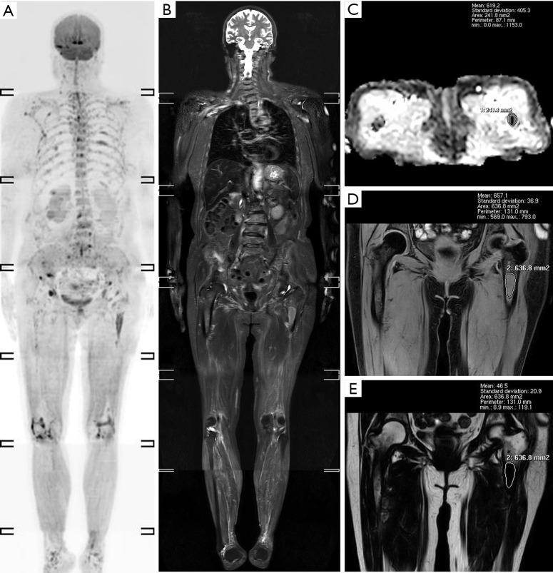
Figure 3.
A 73-year-old female MM patient with a focal infiltration MRI presentation. (A) DWIBS and (B) STIR shows multiple focal signal abnormalities, of which the lesion with the largest diameter is located at the upper segment of the left femur. (C) The ADC map of the left femur’s upper segment shows that the mean ADC value is 0.62×10⁻³ mm2/s. According to the (D) W image and (E) F image in mDIXON, the mean fat fraction of the upper segment of the left femur is 6.6%. STIR, short TI inversion recovery; DWIBS, diffusion-weighted whole-body imaging with background body signal suppression; ADC, apparent diffusion coefficient.

