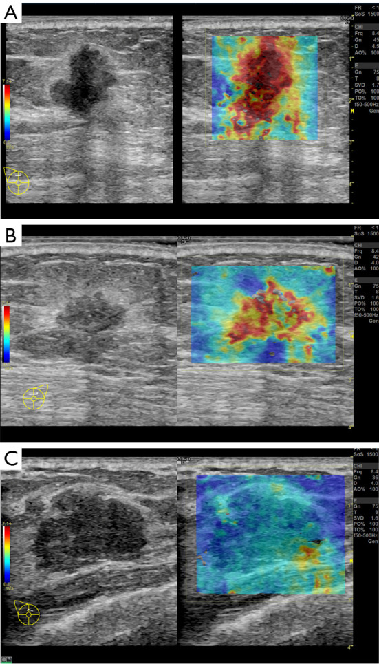
Figure 1.

B-mode (left) and SWE (right) images of breast cancer (A,B) and breast FA (C) in split screen mode. (A) Grade II infiltrating ductal carcinoma. SWE depicts an almost red-colored mass, with an Emean of 149 kPa. (B) Grade II infiltrating ductal carcinoma. SWE depicts a yellow-to-red-colored mass with an Emean of 76 kPa. (C) Breast FA with an Emean of 19 kPa; SWE depicts a blue-to-yellow color for the lesion. SWE, shear wave elastography; FA, fibroadenoma.
