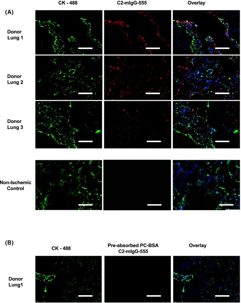
FIGURE 7.
C2 epitope is present in ischemic human donor lungs. A. Immunofluorescent localization of C2 antigen using a chimeric C2-mIgG antibody for detection. Lung tissues were collected from donor lungs (n = 3), trimmed to enable reduced size lobar LTx and lung tissues from non–ischemic control lungs (n = 3, from lung tumor resections). C2 epitope staining with C2-mIgG-Alexa 555 (Red) and Pan cytokeratin (green) used for tissue architecture, demonstrated the presence of the C2 epitope (red) dispersed throughout the lung tissues from ischemic donor lungs, but were not present in nonischemic controls (representative of n = 3). B. Specificity of the C2-mIgG-555 antibody was confirmed by preincubating the antibody with PC-BSA, a known binding epitope of C2, which resulted in loss of C2 epitope staining in the donor lung samples (refer to methods for details). Representative images from 18 samples from six different patients. Scale bar 100 μm

