
Figure 1.
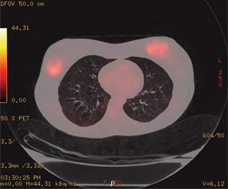
Axial paranchimal fused PET/CT image showing massive lesions of 4 cm in the right breast and 5 cm and 1.5 cm in the left breast (SUVmax = 9) and 1 cm (SUVmax = 3.7) increase in metabolism areas in both axilla
PET/CT: Positron emission tomography/computed tomography