
Figure 2.
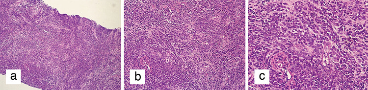
Microscopic images of diffuse blastic cell infiltration in breast tissue with haematoxylin and eosin stain (H&E). a) Diffuse blastic cell infiltration in breast tissue (H&E x100). b) Diffuse blastic cell infiltration in breast tissue (H&E x200). c) Diffuse blastic cell infiltration in breast tissue (H&E x400)