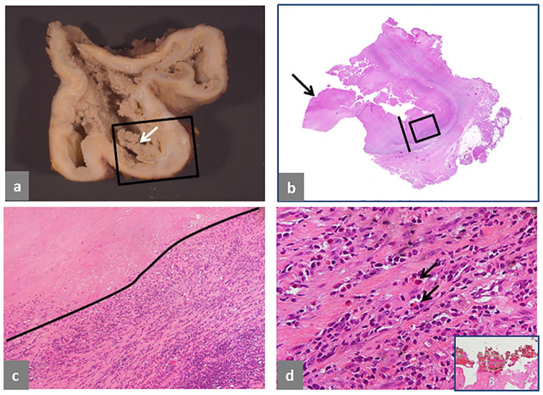
Fig. 3.
Pseudotumor, mixed solid and cystic, extracapsular. (a) Trochanteric bursa, 12 × 10 × 8 cm with necrotic wall and papillary, necrotic neo-synovium (white arrow). (b) Histological section of the area in the black box in a showing wall thickness of 12 mm (black bar) and necrotic papilla (black arrow). (c) Detail of the bursal wall in the black box in b shows tissue necrosis and a large band of predominant lymphocytic infiltrate (H&E × 40). (d) Detail of the inflammatory infiltrate shows presence of numerous eosinophils indicated by black arrows (H&E × 400) and a large aggregate of greenish microplates of metallic corrosion particle aggregates alternating with red layers of blood products in inset (H&E × 200). The case is of a non-MoM THA with CoCr DMN and TMZF stem, implanted for 68 months, with pain in the gluteal area 13 months before revision and pseudotumor identified on MRI study after onset of symptoms. Serum levels of Co and Cr were not performed.
Note. H&E, haematoxylin and eosin; MoM, metal-on-metal; THA, total hip arthroplasty; CoCr, cobalt-chromium; DMN, dual modular neck; TMZF, Ti, Mo, Zr, Fe; MRI, magnetic resonance imaging.

