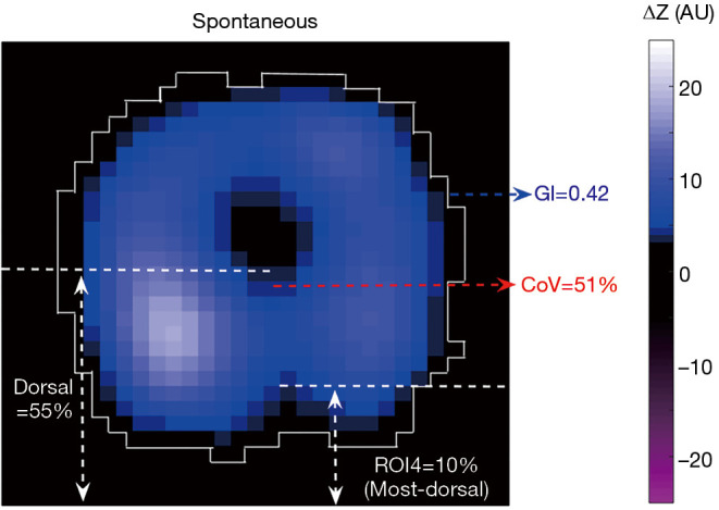
Figure 1.

Illustration of EIT-based parameters in a tidal variation image of an individual with healthy lungs during spontaneous breathing. Regional ventilation delay can only be assessed with a series of EIT-images and is therefore not included in this illustration. EIT, electrical impedance tomography; GI, the global inhomogeneity index; CoV, center of ventilation; ROI4, the 4th region of interest; ΔZ, relative impedance changes; AU, arbitrary unit.
