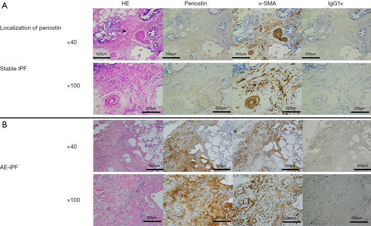
Figure 5.
Representative findings of SLB and autopsied specimens obtained from a 66-year-old man with IPF. (A) Stable IPF. SLB specimens of stable IPF showed strong expression of periostin in fibroblastic foci, with patchy distributions. (B) AE-IPF. Autopsied specimens obtained from patients with AE-IPF also showed strong expression in fibrotic lesions. The numbers of periostin-positive cells and α-SMA-positive cells in fibrotic lesions were higher in AE-IPF than in stable IPF. HE: hematoxylin and eosin staining; Periostin: rat anti-human periostin monoclonal antibodies (mAbs) of immunoglobulin G1κ (IgG1κ; clone no. SS19B or SS5D, produced in the laboratory of Biomolecular Sciences, Saga Medical School, Saga, Japan); α-SMA: mouse anti-human alpha-smooth muscle actin (α-SMA)-Abs (clone 1A4, Dako, Japan); IgGκ: purified mouse IgG1κ-Abs (BioLegend, USA).

