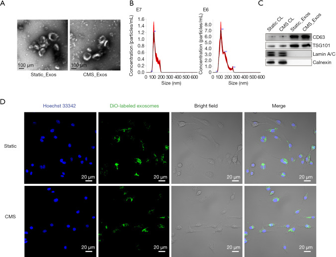
Figure 1.
Characterization of exosomes and incorporation of exosomes into BMMs. (A) Electron microscopy images of exosomes. Scale bar, 100 nm. (B) The size distribution and concentration of exosomes secreted by BMSCs measured by nanoparticle tracking analysis. (C) Western blotting for the exosomal markers CD63 and TSG101, nuclear proteins LaminA/C and the endoplasmic reticulum protein Calnexin. (D) Confocal microscopy images showing the colocalization of exosomes from BMSCs with BMMs. Exosomes were labeled with DiO (green), and BMM cell nuclei were stained with Hoechst 33342 (blue). BMM, bone marrow macrophage; BMSCs, bone marrow mesenchymal stem cells; DiO, 3,3’-dioctadecyloxacarbocyanine perchlorate.

