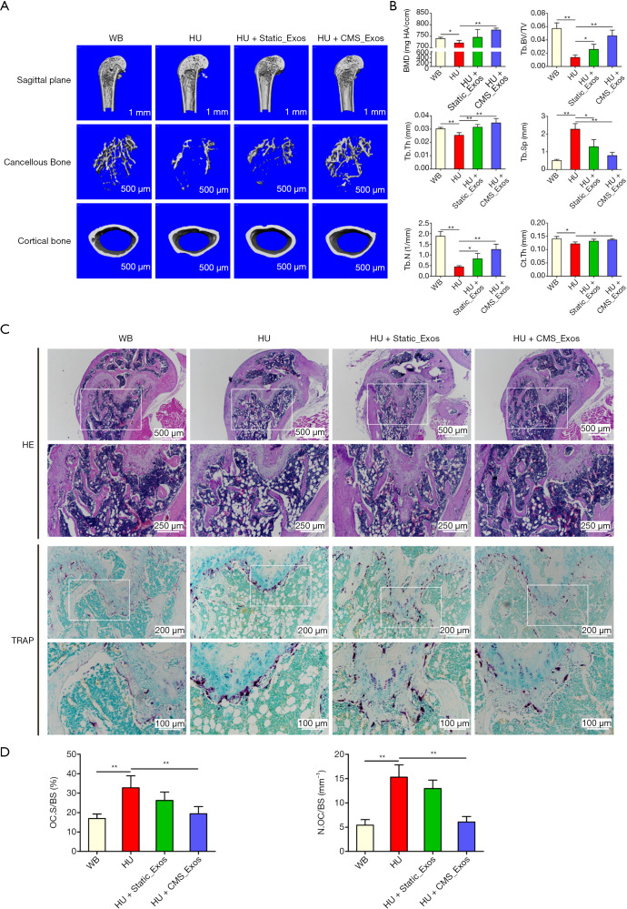
Figure 5.
CMS_Exos prevented HU-induced bone loss. (A) Representative reconstructed micro-CT images of the distal femur in the indicated group. n=5. (B) Three-dimensional microstructural parameters of the distal femur in the indicated group. (C) Representative H&E and TRAP staining images of the distal femur showing the bone volume in the indicated group. n=5. (D) The percentage of osteoclast surface per bone surface (OcS/BS, %) and number of osteoclasts per field of tissue (No. of OCs per field) at 200× magnification were analyzed. Data are expressed as the mean ± SD; *P<0.05, **P<0.01. CMS_Exos, CMS-treated BMSC-derived exosomes; HU, hindlimb unloading; micro-CT, micro computed tomography; H&E, hematoxylin-eosin; TRAP, tartrate-resistant acid phosphatase; SD, standard deviation.

