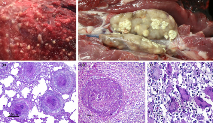
Figure 2.

TB compatible lesions in wild boars. (a) Wild boar, Lung section. Miliary granulomatous pneumonia. This animal was likely excreting numerous mycobacteria. (b) Wild boar, submandibular lymph node section. Multifocal caseous granulomatous lymphadenitis. (c) Wild boar, lung (same as shown in a). Four neighbouring encapsulated granulomas in the lung parenchyma. H&E. Bar 250 μm. (d) Wild boar, submandibular lymph node. Chronic encapsulated granuloma in the lymph node parenchyma, mostly composed of necrosis, mineralization and minimal macrophage inflammatory infiltrate. H&E. Bar 100 μm. (e): Wild boar, submandibular lymph node. Cluster of multinucleated giant cells (Langhan's cells) diffusely infiltrating the lymph node parenchyma. H&E. Bar 25 μm [Colour figure can be viewed at wileyonlinelibrary.com]
