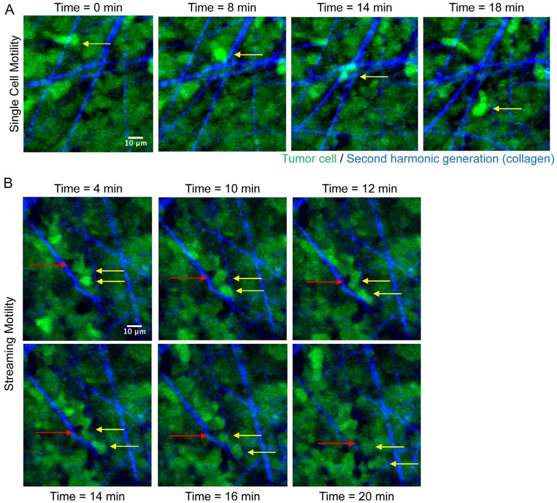
Fig. 4.

Intravital imaging of a melanoma xenograft. Two-photon imaging of tumor cells (in green) and second harmonic generation of fibrillar collagen (in blue). (A) Yellow arrow points to a single cell moving over time (indicated above each panel). (B) Example of streaming motility; yellow arrows point to cancer cells following each other, and red arrow points to another cell type in the tumor microenvironment moving within the multicellular stream. Scale Bar: 10μm. SK-Mel-147 GFP-labeled melanoma cells were injected subcutaneously in 6-week old female nude mice and tumors were allowed to grow up to 1cm3. Intravital imaging of the primary tumor was performed as in Patsialou et al. (2013) and collagen fibers were visualized by second harmonic generation. Images were acquired every 2min for 30min, with a step-size of 5μm.
