Fig. 1.
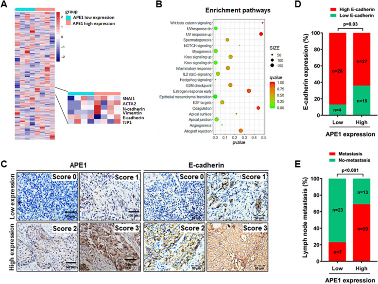
APE1 expression associated with EMT and lymph node metastasis in cervical cancer patients. a Heatmap showing genes differentially expressed in cervical cancer with high expression (n = 4) and low expression of APE1 (n = 4). If the APE1 expression level in cancer tissues was higher than adjacent tissues, it was classified as the APE1 high expression group and if there was no difference, it was classified as the APE1 low expression group. b Gene set enrichment analysis (GSEA) of signaling pathways. GSEA was performed using transcriptome data from cervical cancer samples with high expression (n = 4) and low expression of APE1 (n = 4). If the mRNA expression level of APE1 in cancer tissues was higher than adjacent tissues, it was classified as the APE1 high expression group and if there was no difference, it was classified as the APE1 low expression group. c The expression levels of APE1 and E-cadherin were determined by immunohistochemistry (IHC) of 72 specimens of cervical cancer patients. The representative images are the standard scoring images used to evaluate the intensity of APE1 and E-cadherin staining. d The expression of APE1 and E-cadherin was negatively associated in cervical cancer patients. e A high expression level of APE1 was correlated with lymph node metastasis in cervical cancer patients. The difference was tested by the chi square test

