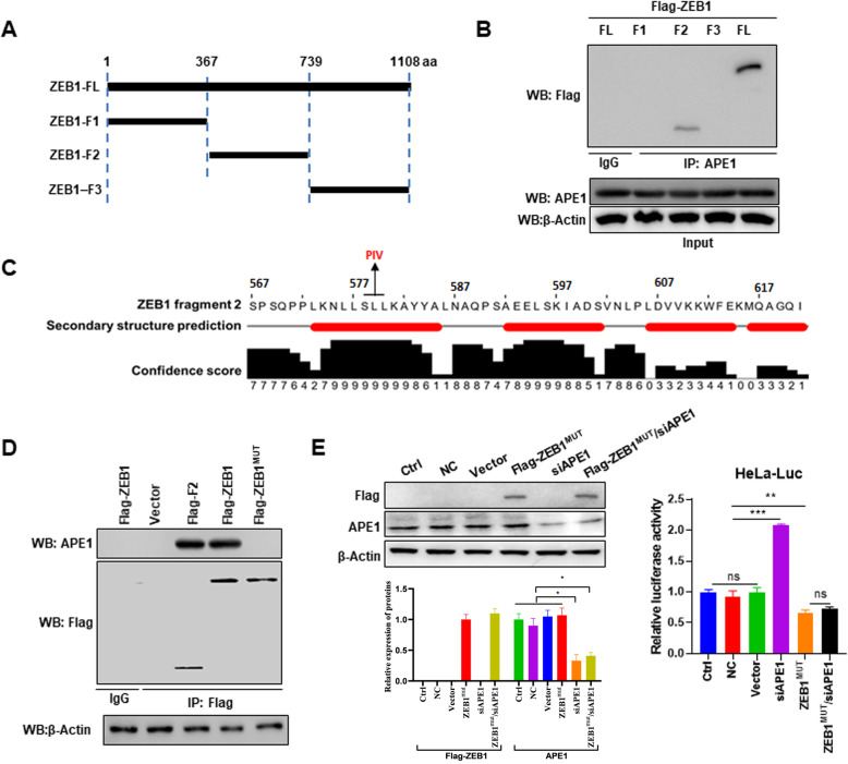
Fig. 5.
ZEB1 amino acids 578–580 are essential for the interaction with APE1. a Schematic representation of ZEB1 protein fragments. b Co-IP showing that the middle fragment of ZEB1 binds to APE1 (F2). Co-IP was performed after 72 h of transfection. c Secondary structure prediction of ZEB1-F2 suggesting the formation of an α helix. d Co-IP showing that mutation of ZEB1 578–580 disrupts the ZEB1-APE1 interaction. Co-IP was performed after 72 h of transfection. e Silencing of APE1 is unable to rescue ZEB1 induced inhibition of luciferase activity in the setting of the ZEB1 578–580 mutation. Western blot and luciferase assays were performed after 72 h of transfection. FL, full-length ZEB1; F1, ZEB1 fragment 1; F2, ZEB1 fragment 2; F3, ZEB1 fragment 3; Vector, cells transfected with empty vector; NC, cells transfected with nontargeting control siRNA; Ctrl, cells not treated with anything. Data are presented as the mean ± SD. ***, P < 0.001

