Figure 5.
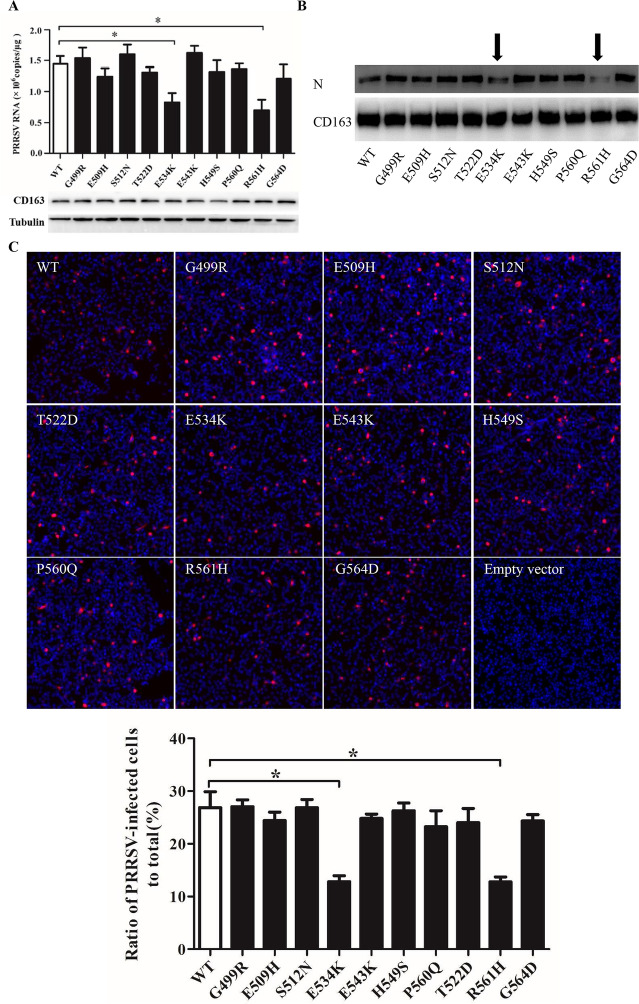
Identification of residues in pCD163 SRCR5 important for PRRSV-2 infection. A RT-qPCR analyses of the effect of mutated pCD163 on PRRSV-2 replication. WT or mutant pCD163 constructs (3 μg/well, 6-well plate) were transfected into PK-15 cells. After 24 h, the transfected PK-15 cells were inoculated with PRRSV-2 strain BJ-4 at a MOI of 1. At 12 hpi, total RNAs of infected PK-15 cells were extracted and then the viral RNA was measured by RT-qPCR. In parallel, expression levels of WT and mutant pCD163 were tested by IB using a commercial mouse anti-human CD163 antibody (MCA1853). Data represent means ± SEM from three independent experiments. *p < 0.05. B Analyses of PRRSV N protein expression in WT or mutant pCD163-expressed cells. The infected WT or mutant pCD163-expressed cells were harvested and lysed at 24 hpi. PRRSV N protein in WT or mutant pCD163-expressed cells was measured by IB. Significant reduction in PRRSV N protein expression is marked by an arrow. In parallel, expression levels of WT and mutant pCD163 were tested by IB using a commercial mouse anti-human CD163 antibody (MCA1853). C PRRSV infectivity in WT or mutant pCD163-expressed cells. The infected cells (24 hpi) were fixed and stained with PRRSV N protein (red) antibody. Nuclei were stained with DAPI and examined by IFA. The total fluorescence intensity of PRRSV N protein was calculated using ImageJ software. Data represent means ± SEM of three independent experiments. *p < 0.05.

