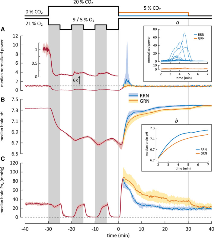
FIGURE 5.

Changes in cortical activity (A) and in brain pH (B) and Po2 (C) in rat pups exposed to intermittent asphyxia, followed by rapid (RRN, n = 17) or graded restoration of normocapnia (GRN, n = 12). All values are median with 95% confidence interval except for insets. The three vertical gray columns on data panels mark the 9% O2 bouts during the intermittent protocol, whereas the two vertical lines indicate the end of asphyxia and the end of GRN. Median data for the RRN and GRN protocols are plotted separately starting at 2 min after the end of asphyxia, as the conditions before this time point are identical in the two protocols. (A) Power of local field potentials (LFPs) in the parietal cortex. The trace with a separate y‐axis is a sixfold magnification of the LFP power and shows the profound suppression of cortical activity during asphyxia (cf. Figure 4). After asphyxia, the activity recovers promptly. Following RRN, but not GRN, a period of hyperexcitability and seizures was seen, and it coincided with the rapid alkaline recovery of brain pH during a time window of 2–7 min after the end of asphyxia. Inset a shows individual LFP power traces during this period. (B) Brain pH decreases by 0.6 pH‐units during the asphyxia and displays only minor modulation (0.12 pH‐units) upon the changes between the 9% and 5% O2 levels. The alkaline recovery of brain pH after RRN is biphasic, and it is slowed down by GRN. The median brain pH during the first recovery phase (2–7 min) is shown magnified in inset b. (C) Brain Po2 falls to apparent zero during the three periods of exposure to 5% O2, but recovers to achieve a normoxic level when the ambient O2 concentration is increased to 9%. After the asphyxia, an overshoot of Po2 is observed. GRN increases the duration but not the amplitude or the rate of the Po2 overshoot
