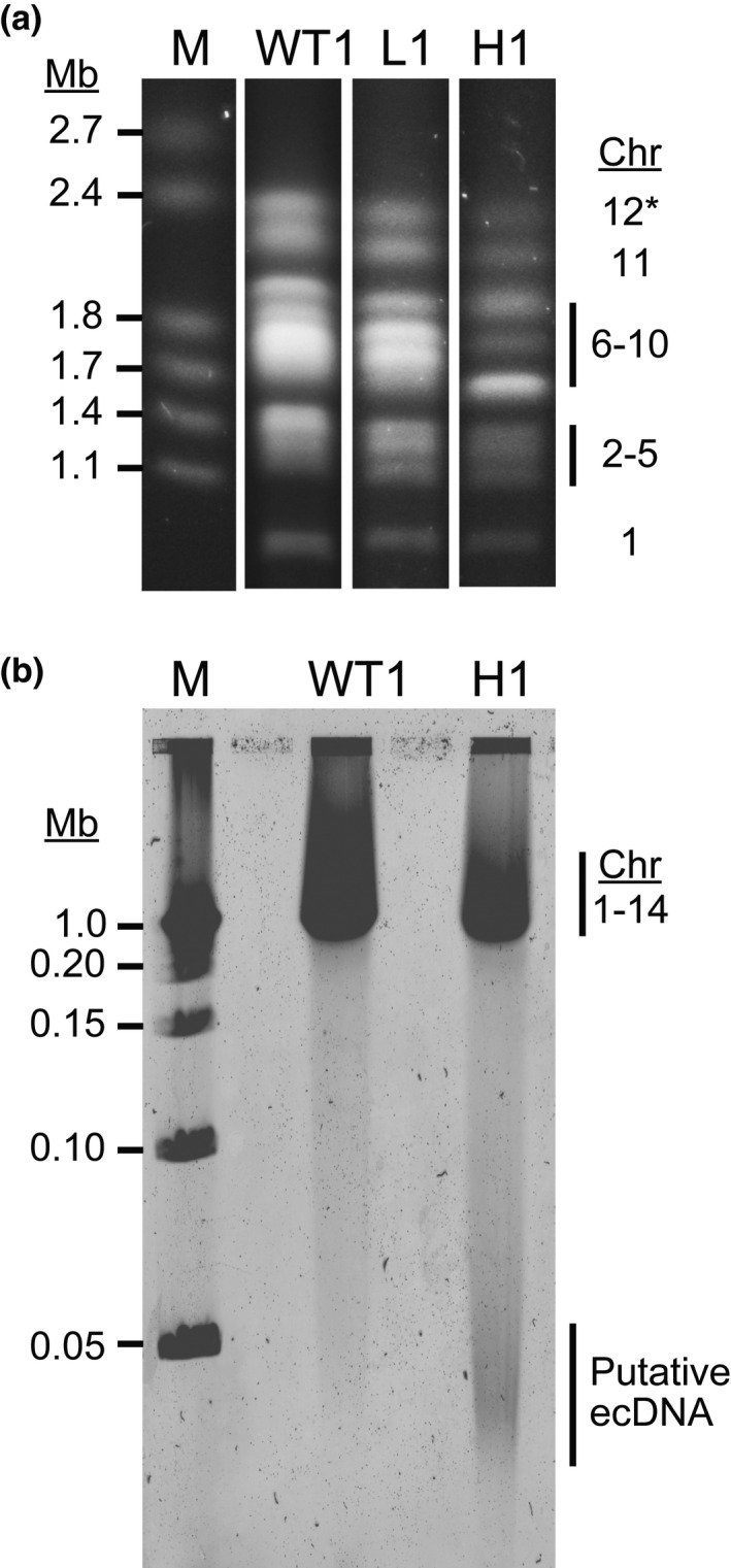
FIGURE 2.

Pulse field gel electrophoresis reveals chromosomal shifts and aberrant DNA elements in highly resistant parasites. (a) Wild‐type (WT1:Dd2), low‐ (L1), and high‐level resistant parasites (H1) were embedded in agarose plugs, run on PFGE (50 hr, 3V/cm, 250–900 sec switch rate), stained with ethidium bromide, and imaged on a UV transilluminator. M, marker (1–3.1 Mb, BioRad 170‐3667). *Although not visualized in this blot, chromosomes 13 and 14 ran above chromosome 12 and appeared as expected in all clones. (b) Parasites were treated as in panel a prior to the PFGE run (17 hr, 6V/cm, 1–10 sec switch rate), stained with SYBR Safe DNA stain, and imaged using the Typhoon 9410 Variable Mode Imager. M, marker (0.05–1 Mb, BioRad 170‐3635); Chr, chromosome
