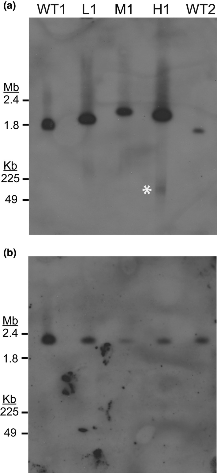
FIGURE 3.

Southern blot analysis detects the resistance gene on both chromosomal and extrachromosomal DNA. Wild‐type (WT1:Dd2, WT2:HB3), low‐ (L1), moderate‐ (M1), and high‐level resistant parasites (H1) were embedded in agarose plugs, run on PFGE (50 hr, 3V/cm, 250–900 sec switch rate), transferred to a membrane, and probed for specific genomic loci. For all blots, the entirety of the gel is shown below the loading well and DNA size was determined with a 1–3.1 Mb marker (upper gel region, Bio‐Rad 170‐3667) and a 0.05–1 Mb marker (lower gel region, Bio‐Rad 170‐3635). (a) Southern blot probed for thedihydroorotate dehydrogenasegene (dhodh, Table 1) that is contained within the amplicon and confers DSM1 resistance (Guler et al.,2013). The expected size of WT1 chromosome 6 is 1.35 Mb and WT2 chromosome 6 is 1.33 Mb (PlasmoDB [Aurrecoechea et al.,2009]). Film exposure time: 8 hr. White asterisk, gel‐competent ecDNA. (b) Southern blot probed for single copy gene on chromosome 11 (Table 1). Analysis of an additional housekeeping gene was also performed on a different blot (single copy gene on chromosome 7, Table 1, FigureS2b). The expected size of WT1 and WT2 chromosome 11 is ~2 Mb (PlasmoDB [Aurrecoechea et al.,2009]). Film exposure time: 7 hr
