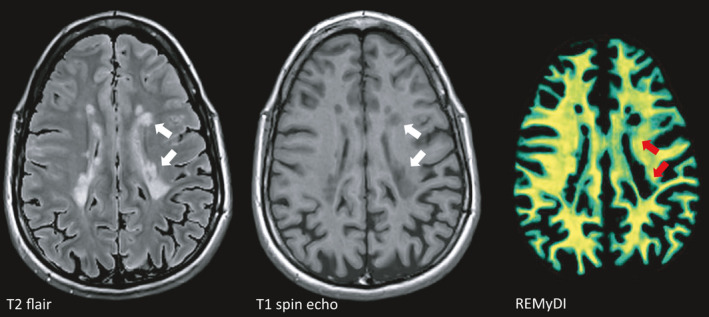
Fig. 2.

Magnetic resonance imaging (MRI) is the most important non‐clinical tool to monitor disease progression. On T2 flair‐weighted images, accumulation of hyperintense (bright) lesions around the ventricles is one of the hallmark signs of MS. These lesions appear hypointense (dark) on T1‐weighted images as a sign of more pronounced tissue destruction. Rapid Estimation of Myelin for Diagnostic Imaging (REMyDI) represents a novel technique to visualize myelin integrity using standard MRI equipment [63]. In this image, complete or near complete loss of myelin is seen in T1 hypointense lesions, but more widespread affection of myelin is evident also in white matter areas outside of focal lesions (green areas as contrasted by yellow areas with intact myelin). Images courtesy of Tobias Granberg, KI, Sweden.
