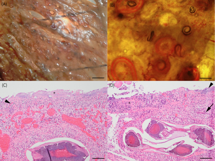
Figure 3.

Intestine from 2‐year‐old horse with acute cyathostominosis (Case 2); Illumination of the mucosal layer of the ventral colon with large numbers of L4 cyathostomins in (A) bar = 2 cm, (B) bar = 1 cm. Photomicrograph of caecum (C) shows diffuse severe mucosal necrosis and loss with infiltration of large numbers of coccoid bacteria (asterisk). There is vascular thrombosis with infiltration of neutrophils (arrowhead). The submucosa is infiltrated by macrophages surrounding the encysted L4 and markedly dilated blood vessels are present. Similar changes are seen in the ventral colon (D) with marked mucosal necrosis (asterisk) and bacterial overgrowth (arrowhead). The submucosa is densely infiltrated by macrophages surrounding L3 (arrow) and encysted L4. Haematoxylin and Eosin stain, bar = 100 µm
