Fig. 2.

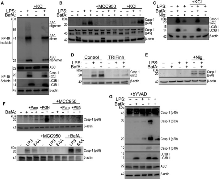
NLRP3 dependence of the V‐ATPase effect in monocytes. (A) Western blots of DSS cross‐linked NP‐40 insoluble ASC oligomers and NP‐40 soluble ASC, caspase‐1 (p20), and LC3B from CD14+ monocyte total cell lysates stimulated with LPS (1 µg·mL−1) plus and minus bafilomycin A1 (BafA, 100 nm) and extracellular KCl (20 mm) for 18 h (n = 4). (B) Western blot of mature caspase‐1 (p20) from CD14 + monocyte total cell lysates stimulated with LPS (1 µg·mL−1) plus and minus BafA (100 nm) supplemented with MCC950 (5 µm) or extracellular KCl (20 mm) (n = 3). (C) Western blot of mature caspase‐1 (p20) from CD14+ monocyte total cell lysate stimulated with LPS (1 µg·mL−1) plus and minus BafA (100 nm) or nigericin (Nig, 10 µm) supplemented with extracellular KCl (20 mm) for 18 h. Nigericin was added at the last hour of stimulation (n = 2). (D) Western blot of caspase‐1 (p20) from CD14 + monocyte total cell lysate treated with control peptide or TRIF blocking peptide (TRIFinh, 50 µm) for 2 h, before stimulation with LPS (1 µg·mL−1) plus and minus BafA (100 nm) for 18 h (n = 4). (E) Western blot of caspase‐1 (p20) from naïve or LPS‐primed (1 µg·mL−1, 4 h) THP‐1 monocyte total cell lysates preincubated with or without BafA (100 nm, 15 min) before addition of nigericin (10 µm, 1 h) (n = 4). (F) Upper panel: Western blot of mature caspase‐1 (p20), and β‐actin, from CD14 + monocyte total cell lysate stimulated with Pam3CSK4 (Pam, 10 μg·mL−1), or PGN from S. aureus (PGN, 20 μg·mL−1) plus or minus bafilomycin A1 (BafA, 100 nm) and/or MCC950 (10 μm) for 18 h (n = 3). Lower panel: Western blot of mature caspase‐1 (p20), and β‐actin, from CD14+ monocyte total cell lysate stimulated with LPS (1 μg·mL−1) or SAA (5 μg·mL−1) plus or minus MCC950 (10 μm) and BafA (100 nm) for 18 h (n = 2). (G) Western blot of caspase‐1 p45, p33, p20 and p10 species, and lipidated LC3B detected from CD14+ monocyte total lysates stimulated with LPS (1 µg·mL−1) plus and minus BafA, (100 nm) or biotin‐YVAD‐cmk (bVAD, 50 µm) for 18 h (n = 5).
