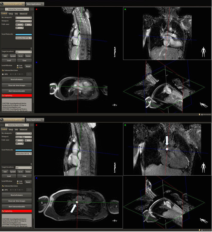
FIGURE 3.

Images acquired with iSuite in a patient with Tetralogy of Fallot and LPA stent. An increase in the pSAT angle from 30° (top image) to 45° (bottom image) improved the conspicuity of the balloon‐tip by increasing the contrast between blood and balloon.
