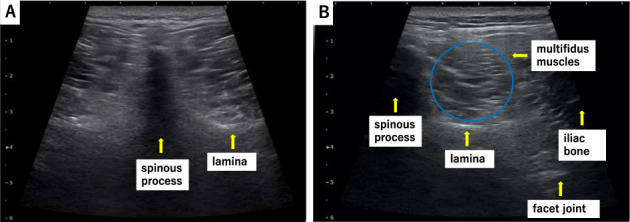
Figure 1.

The multifidus muscle and surrounding anatomy at the L4/5 level in a healthy volunteer. (A) A probe is applied vertically to identify the spinous process on the axial view and (B) moved to the right or left to identify each tissue area of interest.
