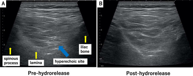
Figure 2.

Hydrorelease of the multifidus muscle. Hydrorelease was performed using 7.0 mL of saline in the hyperechoic site of the multifidus muscle (A) pre‐hydrorelease; the blue arrow indicates the hyperechoic site. (B) Post‐hydrorelease, the hyperechoic site has disappeared.
