FIGURE 3.

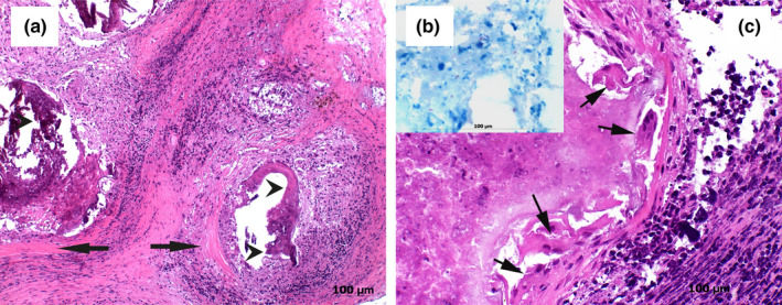
Histological findings from a mandibular lymph node presenting histological lesions compatible with tuberculosis and tested positive for Mycobacterium microti. (a) Overview of a granulomatous lymphadenitis showing focal‐extensive necrotic cores and dystrophic calcifications (arrow heads). The necrotic cores are surrounded by a mixed population of inflammatory cells and by a wide capsule of connective tissue (arrows), haematoxylin and eosin (HE; 40×). (b) Scanty acid‐fast rods are visible in the necrotic debris. Occasionally, S‐shaped bacilli, which are commonly associated with Mycobacterium microti were observed intra‐ and extracellularly, Ziehl‐Neelsen stain (40×). (c) Granuloma characterized by focal‐extensive necrosis and mild inflammatory infiltration of epithelioid macrophages, neutrophils, multinucleated Langhans type giant cells (arrows) and eosinophils is shown (HE; 40×)
