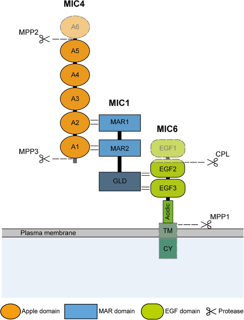
FIGURE 1.
Schematic representation of domain structure and hydrolysis modification of TgMIC1/4/6. The domain structure and interaction sites of TgMIC1/4/6 are shown. And the C-terminal galectin-like domain (GLD) of MIC1, transmembrane (TM) and cytoplasmic domain (CY) of TgMIC6 are also indicated. TgMIC4 and TgMIC6 undergo hydrolysis modification by MPP2, MPP3, and CPL, respectively, during transport to the microneme, and then the complex is anchored on the surface of the parasites by TgMIC6, and finally released by MPP1 after the invasion is completed. The cleavage sites are all plotted.

