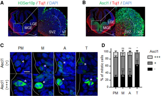
Figure 2.
Quantification of Ascl1 expression in apically dividing neural progenitors of ventral telencephalon. (A,B) Cross-sections of mouse telencephalon immunostained for pHH3 and Tuj1 (A) or Ascl1 and Tuj1 (B) together with DAPI staining. Mitotic pHH3+ cells can be found in the ventricular zone (VZ) and subventricular zone (SVZ) in both the lateral and medial ganglionic eminences (LGE and MGE, respectively) of E12.5 ventral telencephalon. (C) The cell cycle stage of apically dividing progenitors, both from LGE and MGE, was assessed using DAPI staining and segregated in cells not expressing (−), expressing low levels of (+), or expressing high levels of (+++) Ascl1 protein. (D) Stacked bar plots showing the percentage of mitotic cells expressing different levels of Ascl1 in prometaphase (PM), metaphase (M), anaphase (A), and telophase (T). Data shown as mean percentage ± SD. Data information: Cross-sections from three mice were used. From each mouse, six consecutive slices were acquired in a total of 1736 cells being quantified. One-way ANOVA Tukey's multiple comparison was used to compare Ascl1 levels between prometaphase and other M-phase stages. (ns) P > 0.05, (*) P ≤ 0.05, (**) P ≤ 0.01. Scale bars:, A,B, 100 µm; C, 5 µm.

