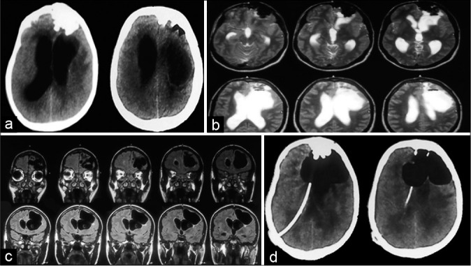
Figure 1:

(a) Plain computed tomography (CT) brain showing left frontal irregular bony growth with intracranial projection, ventriculomegaly and left paraventricular cystic SOL. (b) Magnetic resonance imaging (MRI) brain plain T2 axial images showing irregular hypointense left frontal lesion encroaching on left frontal lobe, paraventricular cystic lesion of cerebrospinal fluid intensity is noted with air-fluid level in it. (c) T1 coronal MRI depicting relation of cyst to ventricle. (d) Post-op CT brain plain following ventriculoperitoneal shunt done for hydrocephalus showing enlargement of aerocele.
