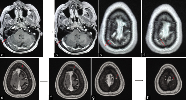
Figure 3:
Illustrations of the different pathways for the diploic veins. The MRI in (a and b) shows a diploic vein in the right occipital region marked with red arrow appearing to drain directly into the transvers sinus. The MRI in (c and d) shows diploic veins marked with red arrow in the right parietal region which appear to drain into the lateral aspect of the superior sagittal sinus. The MRI in (e and f) shows a diploic vein in the left frontal region marked with red arrow appearing to drain into the superior sagittal sinus. The MRI in (g and h) shows a diploic vein in the left parietal region marked with red arrow appearing to drain into the lacunae laterales.

