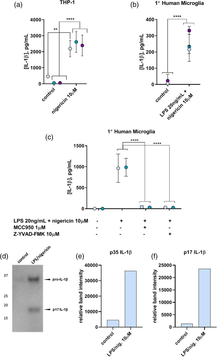
FIGURE 1.

(a) IL‐1β in culture supernatants of PMA‐differentiated THP‐1 cells upon canonical inflammasome activation with nigericin alone (10 μM, 30 min) in the absence of LPS priming, as measured by ELISA. (b) IL‐1β in culture supernatants of primary human microglia upon LPS priming (20 ng/ml, 3.5 hr) followed by nigericin activation (10 μM, 30 min). (c) Inhibition of canonical NLRP3 inflammasome activation in primary human microglia by inhibiting NLRP3 with MCC950 or caspase‐1 activity with Z‐YVAD‐FMK. ** = p = .001; 2 or 3 experiments, n = 3. Colors indicate individual experiments. (d) IL‐1β western blot of combined, concentrated supernatants from several primary human microglia experiments showing secretion of p35 pro‐IL‐1β and the mature p17 form upon canonical inflammasome activation with LPS priming (20 ng/ml, 3.5 hr) followed by nigericin activation (10 μM, 30 min). (e,f) Quantification of relative p35 (e) and p17 (f) IL‐1β band intensities between no treatment (control) and canonical inflammasome activation in primary human microglia [Color figure can be viewed at wileyonlinelibrary.com]
