
FIGURE 4.
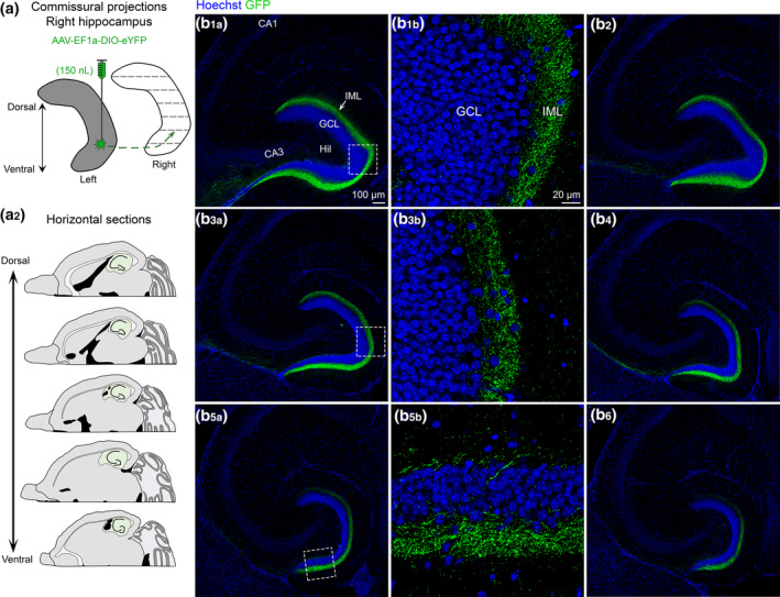
Contralateral projections of ventral MC axons across the septotemporal axis of the DG. (a1) To evaluate contralateral projections of ventral MCs, the left hilus was injected with AAV‐EF1a‐DIO‐eYFP (grey) and the right hippocampus (white) was evaluated in the horizontal plane. (a2) Representative schematic of horizontal sections from dorsal to more ventral hippocampus (green). (b) A representative example of contralateral GFP+ expression in a female Drd2‐Cre+/− mouse. Sections begin at dorsal levels and progress toward more ventral locations. (b1–b6) The contralateral projections of ventral MCs appear to be primarily restricted to the IML across all sections. HIL, hilus; GCL, granule cell layer; IML, inner molecular layer [Color figure can be viewed at wileyonlinelibrary.com]