
FIGURE 6.
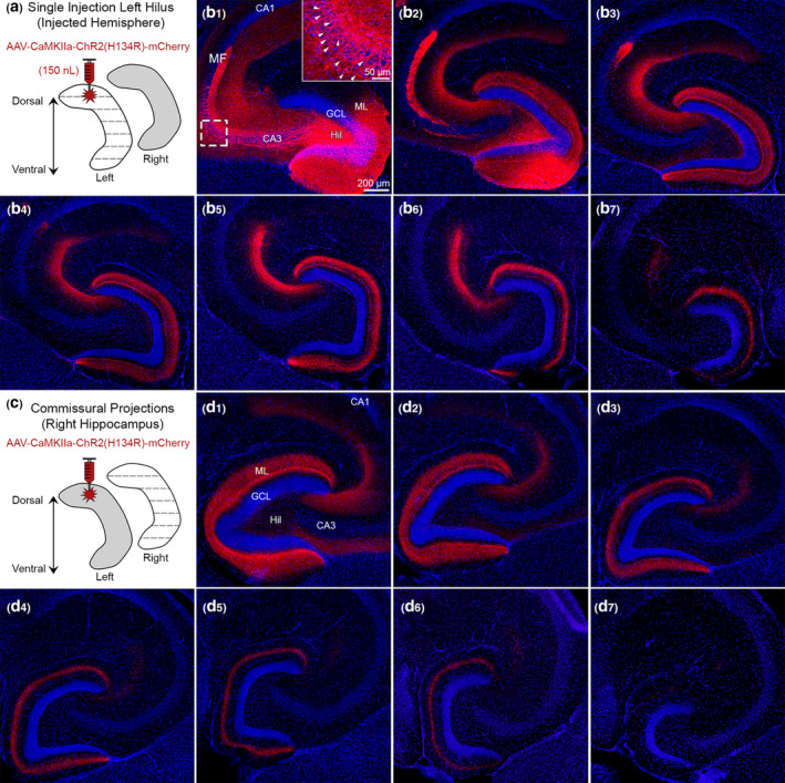
Use of CaMKIIa to probe the specificity of GFP for MCs. (a) Viral injection schematic. A total of 150 nl of AAV‐CaMKIIa‐ChR2(H134R)‐mCherry was injected into the left dorsal hilus to target excitatory neurons. (b1–b2) Near the injection site, viral expression was observed in GCs, MCs, and CA3 pyramidal neurons (inset; white arrowheads). Granule cell mossy fibers (MF) axons were also labeled where they normally project, CA3 stratum lucidum. (b3–b7) Long‐range mCherry+ axons showed a similar pattern of viral expression in the molecular layer as Drd2‐Cre or Crlr‐Cre mice injected in the dorsal DG with a virus to express GFP. (c) Contralateral axons were evaluated in the right hippocampus. (d1–d7) Contralateral mCherry+ axons showed a similar pattern in the molecular layer as Drd2‐Cre and Crlr‐Cre injected with a virus expressing GFP in the dorsal hilus. This figure shows that injection of AAV to express CaMKIIa in the dorsal hilus results in a similar pattern of axon labeling as an injection of AAV to express GFP in MCs. Representative images are from a female Drd2‐Cre−/− mouse. GCL, granule cell layer; HIL, hilus; ML, molecular layer [Color figure can be viewed at wileyonlinelibrary.com]