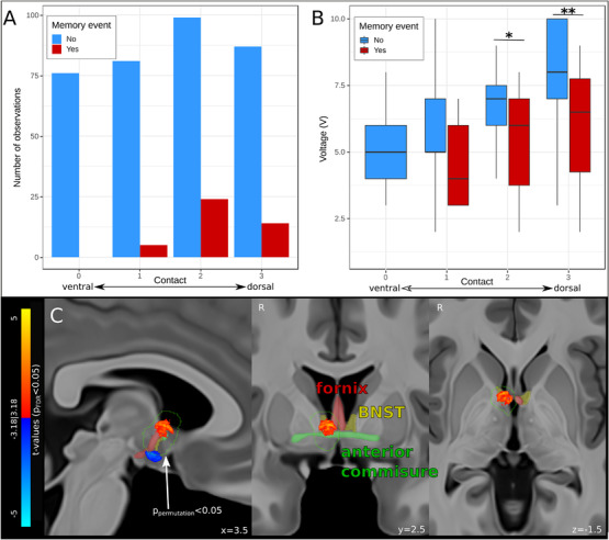
FIGURE 2.

Results of voxel‐wise VTA mapping of flashback‐inducing stimulation. (A) Bar graph showing the number of stimulations (count) at each contact (ventral to dorsal, “0′” to “3′”) that did (red) or did not (blue) produce acute memory events. Note that no memory events were reported during stimulation of the ventral‐most contact. (B) Boxplot showing the minimal voltage (memory event, red) and maximal voltage (no memory event, blue) for the stimulations at each contact. (C) Results of voxel‐wise VTA mapping show voxels significantly (P FDR < 0.05) positively (warm colors) and negatively (cool colors) associated with memory flashbacks. Only the dorsal cluster of significant voxels lay within a region (outlined in green) established as non‐random by non‐parametric permutation testing (P permutation < 0.05, n = 10,000 permutations). The fornix (red), BNST (yellow), and anterior commissure (green) are overlaid in faded colors on sagittal (left), coronal (middle), and axial (right) T1‐weighted MNI152 brain slices. BNST = bed nucleus of stria terminalis; FDR = false discovery rate; MNI = Montreal Neurological Institute; VTA = volume of tissue activated; *P < 0.05; **P < 0.01
