Figure 2.

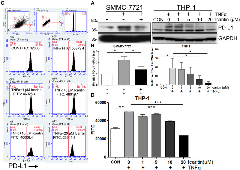
Icaritin inhibits PD‐L1 expression. Cells were treated with 10 μM (SMMC‐7721) or indicated concentrations (THP‐1) of Icaritin for 1 h, followed by 50 ng/mL TNF‐α for 24 h (mRNA level) or 48 h (protein level). The level of PD‐L1 was determined by western blotting, GAPDH was used as the loading control. Blots are representative of three independent experiments (A) or real‐time RT‐qPCR (normalized to GAPDH) (B). (C and D) THP‐1 cells were pretreated with the indicated concentrations of Icaritin for 1 h, followed by 50 ng/mL TNF‐α treatment for 48 h. The level of PD‐L1 was determined by flow cytometry. Gating strategy used to identify THP‐1 cells‐expressing PD‐L1 on their plasma membrane. A first gate was set in living cells, then on physical parameters (FSC‐A vs FSC‐H) to eliminate doublets, and then on PD‐L1+ cells. Data are from one single experiment representative of three independent experiments, three samples are included in each experiment. Values are mean ± SEM (n = 3), *p < 0.05, **p < 0.01, ***p < 0.001 (by Student's t‐test).
