
Figure 5.
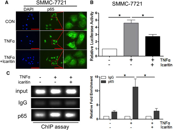
Icaritin inhibits NF‐κB p65 translocation and decrease p65 occupancy on PD‐L1 promoter. (A) Confocal microscopy image showing p65 localization after pretreatment with 20 μM Icaritin for 4 h, followed by treatment with 15 ng/mL TNF‐α for 0.5 h in SMMC‐7721 cells. One representative image from three independent experiments is shown. Scale bar represents 100 μm. (B) Luciferase activity was measured and normalized according to Renilla luciferase activity in SMMC‐7721 cells transiently transfected with the PD‐L1 luciferase promoter (PD‐L1‐Luc). Cells were pretreated with the indicated concentration of Icaritin for 1 h, followed by treatment with TNF‐α for 24 h. (C) Soluble chromatin from SMMC‐7721 cells was precipitated with anti‐NF‐κB p65 or control IgG. The final DNA samples from input or pull‐down group were amplified via qPCR with primers for the PD‐L1 promoter p65‐binding region, data were normalized to input. Each group of cells with three replications were pretreated with 20 μM Icaritin for 12 h, followed by treatment with 50 ng/mL TNFα for 0.5 h. Data are from three independent experiments. Values are mean ± SEM (n = 3). *p < 0.05, **p < 0.01, ***p < 0.001 (by Student's t‐test).