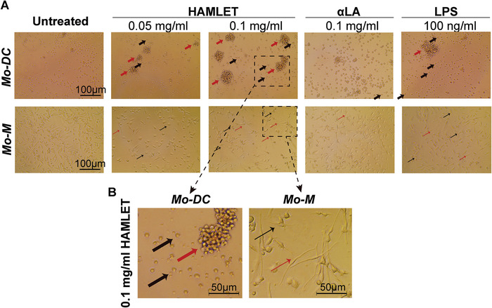
Figure 1.

HAMLET induces morphological changes of Mo‐DCs and Mo‐M. (A‐B) Light microscopy images of primary human Mo‐DCs (upper panel) or Mo‐M (lower panel), treated or not with HAMLET, αLA or LPS at indicated concentrations. After 1‐h of treatment, the medium was replaced with fresh differentiation medium and the cells were grown overnight. Thick black arrows indicate Mo‐DCs with short protrusions and thick red arrows indicate aggregates of Mo‐DCs. Thin black arrows indicate Mo‐M with short protrusions and thin red arrows indicate Mo‐M with elongated morphology. (B) Higher magnification (dashed boxes in A) of Mo‐DCs (left panel) and Mo‐M (right panel) treated with 0.1 mg/ml HAMLET. All images were equally corrected for brightness to improve clarity. Images representative of >3 experiments (n = 1‐2 donors per experiment).
