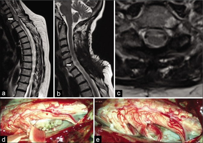
Figure 6:

(a) Preoperative sagittal T2 MRI – ventrolateral meningioma at the level of C7 vertebra (white arrow); (b and c) postoperative sagittal and axial MRI confirms total tumor removal; (d and e) intraoperative images at the beginning and the end of the surgical procedure.
