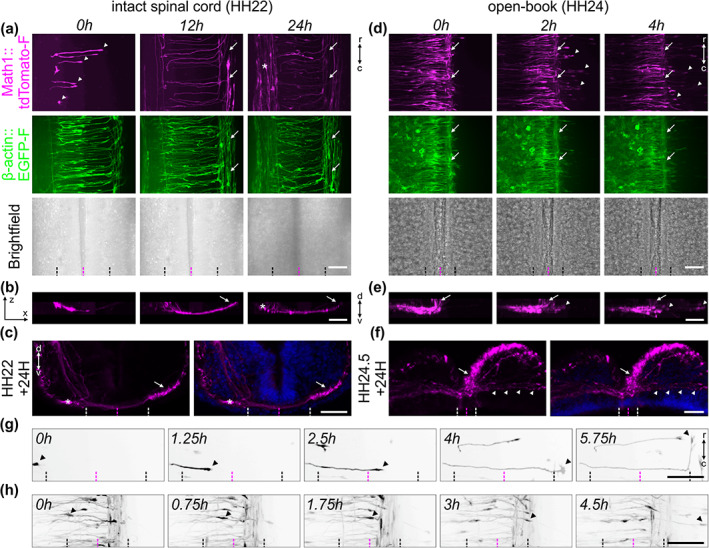
FIGURE 3.

Live imaging of cultured intact spinal cords allowed for the visualization of dI1 axons during floor plate crossing and navigation into the longitudinal axis. (a) 24‐h time‐lapse recording showed that Math1‐positive dI1 commissural axons could cross the floor plate (FP) (white arrowheads), turn anteriorly, as expected, and form the contralateral ventral funiculus (white arrows) in cultured intact HH22 spinal cords. The asterisk indicates a population of Math1‐positive ipsilateral axons. (b) Transversal view of a region of interest from the time‐lapse recording shown in (a), highlighting the trajectory of dI1 axons and the formation of the commissure. The white arrow indicates the position of the contralateral ventral funiculus. The white asterisk labels ipsilateral axons. (c) The normal development of the commissure could be verified by immunochemistry on transverse sections of spinal cords cultured for 24 h. Nuclei were counterstained with Hoechst (shown in blue). (d) 4‐h time‐lapse recording showing Math1‐positive dI1 commissural axons in a cultured open‐book preparation of a HH24 spinal cord. Note that within less than 2 h in culture the majority of dI1 commissural axons were overshooting the contralateral FP boundary and growing straight into the contralateral side after having crossed the FP (white arrowheads). White arrows indicate the contralateral ventral funiculus. (e) Transversal view of a region of interest from the time‐lapse sequence shown in (d) highlighting the aberrant trajectory of dI1 commissural axons (white arrowheads) growing straight past the contralateral ventral funiculus (white arrow). (f) The aberrant development of the commissure could be verified by immunochemistry on transverse sections of open‐books cultured for 24 h. This clearly showed dI1 axons growing straight after having crossed the FP (white arrowheads). Nuclei were counterstained with Hoechst (shown in blue). (g) Single dI1 growth cones (black arrowheads) could be tracked crossing the FP, exiting it and turning rostrally in an intact spinal cord preparation. Math1‐positive axons are now shown in black. (h) Single dI1 growth cones could also be tracked crossing the FP, exiting it, but instead of turning rostrally, most of them overshot in the contralateral part (black arrowheads). Black and white dashed lines indicate FP boundaries, magenta dashed lines the midline, respectively. d, dorsal; v, ventral; r, rostral; c, caudal. Scale bars: 50 μm
