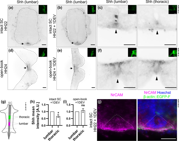
FIGURE 5.

A Shh gradient is still present in cultured intact spinal cords (SCs) but not open‐books after 1 day ex vivo. Intact HH22 SCs or open‐books were cultured for 1 day ex vivo before fixation. Transverse cryosections were immunostained with antibodies against Shh (5E1 clone) and counterstained with Hoechst. (a,b) Shh was still expressed in the floor plate (FP) of intact SCs after 1 day ex vivo in a similar manner as right after dissection (black arrows). (c) Moreover, in agreement with previous descriptions in vivo, Shh was expressed in a decreasing caudal‐to‐rostral gradient with higher expression at the lumbar compared to the thoracic level (black arrowheads). (d,e) Shh was still expressed in the FP of cultured open‐books (black arrows). (f) However, the gradient was lost with no more obvious difference in expression between lumbar and thoracic levels (black arrows). (g) Cartoon depicting the different levels taken for the quantification in (h) and (i). (h,i) Quantification of the mean intensity of Shh staining in the lumbar versus thoracic FP of cultured intact SCs and open‐books. Note that values were normalized to the lumbar level (value = 1). (h) There was a significant decrease in the intensity of Shh in the FP of intact SC at thoracic levels (0.7 ± 0.1) compared to lumbar levels (N = 4 embryos). (i) However, in cultured open‐books this difference disappeared (average value in thoracic level = 1.1 ± 0.1, mean ± SD, N = 4 embryos, two‐tailed Mann–Whitney U test). Error bars represent SD. p < .05 (*) and p ≥ .05 (ns). (j) NrCAM (shown in magenta) was still expressed in the FP (dashed line) of a cultured intact SC. Note that small images on the upper right corner of the different panels show the β‐actin::EGFP‐F‐electroporated side of the SC. The dashed black lines represent the outer border of the SC. d, dorsal; v, ventral; r, rostral; c, caudal. Scale bars: 50 μm
