FIGURE 6.

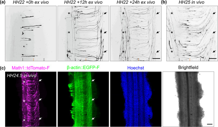
Development of Math1‐positive axonal tracts ex vivo and in vivo. (a) Sequence of three images showing dI1 axons crossing the floor plate (FP) after 0, 12, and 24 h of culture. After turning rostrally, postcrossing axons started to form the contralateral ventral funiculus (black arrows). After around 12 h in culture, a Math1‐positive ipsilateral population could be clearly seen in the ipsilateral ventral funiculus (black asterisks). (b) Intact spinal cords dissected at HH25 (approximately 1 day after HH22), fixed and mounted similarly to the ex vivo culture were imaged the same way. This revealed identical dI1 axonal tracts compared to those seen after 24 h of culture of intact spinal cords dissected at HH22, with postcrossing axons forming the contralateral ventral funiculus (black arrows) and ipsilateral axons turning in the ipsilateral ventral funiculus (black asterisks). Black and orange dashed lines represent FP boundaries and midline, respectively. (c) Low magnification overview of an intact HH24.5 spinal cord, fixed, stained for RFP (Math1‐positive neurons) and GFP (β‐actin transfected cells) and counterstained with Hoechst showing the contralateral ventral funiculus containing postcrossing dI1 axons (white arrows) and the ipsilateral ventral funiculus containing a population of ipsilateral axons (white asterisks). Scale bars: 50 μm (a,b) and 100 μm (c)
