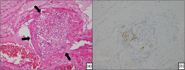
Figure 3.

A representative case of amniotic fluid embolism in an autopsied uterus. (a) Thrombus around the fetal component in a venule of the uterus (arrow). The image shows intravascular coagulation due to the fetal debris. (b) The serial section of Fig. 3a. The fetal debris in the venules of the uterus show positive staining with the broad‐spectrum anti‐pancytokeratin cocktail (AE1/AE3).
