Figure 1.

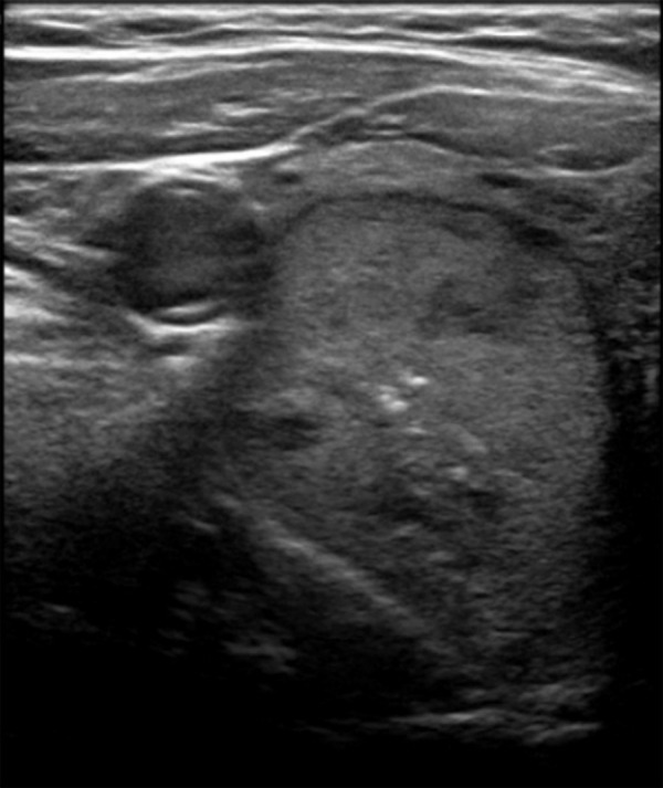
Axial ultrasound image showing a predominantly solid, isoechoic nodule in the right lobe with central punctate echogenic foci which was more difficult to classify using BTA. BTA, British Thyroid Association

Axial ultrasound image showing a predominantly solid, isoechoic nodule in the right lobe with central punctate echogenic foci which was more difficult to classify using BTA. BTA, British Thyroid Association