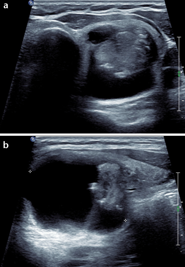
Figure 2.

Axial (a) and longitudinal (b) ultrasound of a papillary carcinoma in the left lobe: a well-defined mixed composition nodule with isoechoic solid component however the solid focus is eccentrically positioned forming a mural nodule that has an acute angle with the cyst wall.
