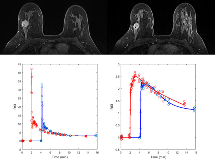
Figure 2.
A 37-year-old female with a Grade 3 triple negative invasive ductal carcinoma in the right breast. T1 weighted HSR images (top) highlight a tumour at baseline (left) with a volume of 3.1 cm3 which reduced to 2.9 cm3 after one cycle of NACT (right). Analysis of the DCE-MRI data using arterial input functions measured in the descending aorta (bottom left) produced estimates of blood flow of 0.46 ml/min/ml tissue at baseline (blue data points and curves) and 0.31 ml/min/ml tissue after one cycle (red data points and curves, bottom right). Following completion of NACT, the patient underwent a wide local excision and pathological assessment of the resected specimen revealed an RCB index of 0 (RCB class 0, pR).

