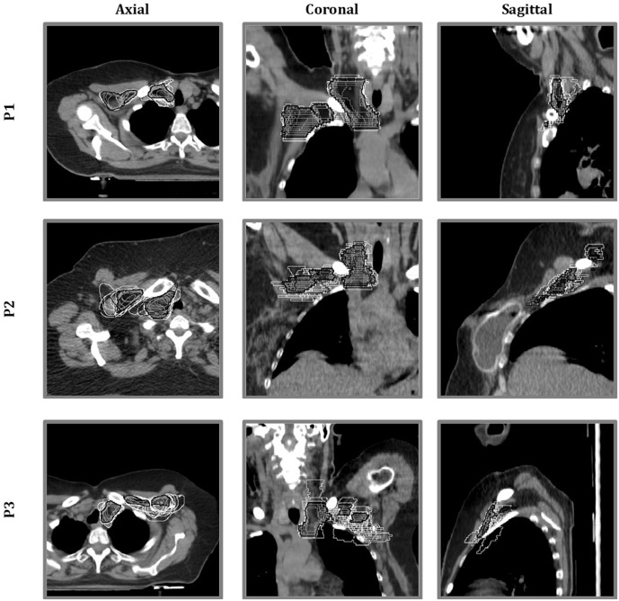
Figure 1.
Qualitative assessment of areas of variability: for all the three representative patients (P1, P2 and P3), all clinical target volumes (CTVs) (single-centre CTVs in white and gold standard CTV in black and grey-filled) were overlaid in the axial, coronal and sagittal CT planes, to visually quantify the interobserver variability.

