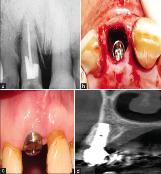
Figure 1.

(a) The preoperative radiograph of a hopeless maxillary central incisor. (b) The osteotomy was prepared palatally so that after implant insertion a large buccal gap remained. (c) At 3 months, excellent soft tissue healing was observed. (d) Cone beam computed tomography images after implant restoration showed reformation of a thick buccal plate of bone.
