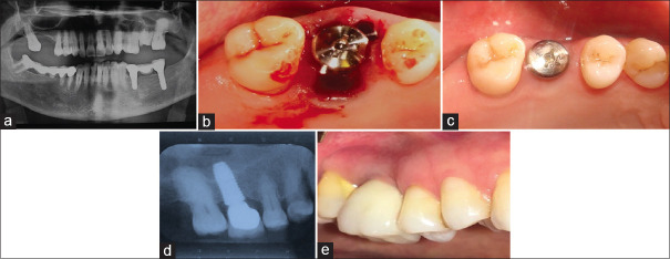
Figure 2.
(a) This patient presented with a hopeless maxillary right first molar tooth. (b) While the osteotomy was originally started in the inter-septal bone, in the end the implant ended up too far buccal leaving only a palatal gap. A 6 mm diameter healing abutment was added, but as a flap-less approach was used, no sutures were needed. (c) After 3 months' site healing, it is apparent that the implant had been positioned too far buccally, and while the palatal gap had healed well, there was a loss of buccal bone resulting in a collapse of the buccal ridge anatomy. (d) At the 1-year follow-up visit, excellent bone healing can be seen. (e) This clinical photo taken at 1 year clearly shows that there has been a collapse of the buccal tissues making the implant suspect for the long-term.

